
Understanding Medical Imaging: A Journey Inside the Black Box
- Andrew Ivanchenko M.D.

- Jan 21
- 4 min read
Updated: Apr 3
The Black Box of the Human Body
Relax—when I refer to the black box, I'm not talking about a coffin. This term describes a system with an unknown design. For a long time, the human body was a black box. We could only observe its contents during an autopsy or surgery, which, as you can imagine, wasn't particularly useful for diagnosing illnesses.
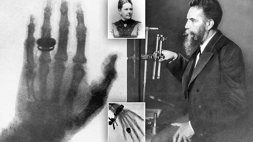
A First Look into the Black Box
Doctors have always dreamed of peeking inside this black box to diagnose illnesses before it was too late. By the end of the 19th century, those dreams began to materialize. The famous Konrad Roentgen, a German physicist, discovered X-rays. These mysterious rays could penetrate the human body, allowing us to visualize bones and, to some extent, internal organs.
X-ray imaging quickly became one of the most popular diagnostic tools. It’s convenient, simple, and inexpensive. X-rays can easily display bone fractures, dark patches in the lungs, or gut perforations. However, many problems remain invisible. An X-ray image is merely a two-dimensional shadow of a three-dimensional object. It takes a lot of imagination to understand what is truly happening inside the body from those overlapping shadows of varying density.
Advancements in Imaging: Computer Tomography
Later in the 20th century, radiologists began using computer tomography (CT). The term "tomo" means "slice" or "layer" in Greek. CT produces images of multiple "slices" of the human body, which are then merged into a single image by a computer. This method allows us to visualize many internal organs and their pathological changes, such as tumors or hemorrhages.
Looking at a CT image feels like traveling inside the human body. Internal organs appear three-dimensional, making it easier to understand their interactions. This breakthrough vastly expanded the scope of accurate diagnostics. Surgeons can now know what to expect during an operation, facilitating their job and reducing the chances of complications.

Unfortunately, both X-ray and computer imaging share a common flaw: they involve radiation exposure. Although the exposure is minimal, it still poses a potential hazard for both the doctor and the patient, especially if the procedure needs to be repeated.
Magnetic Resonance Imaging: A Safe Examination Technique
Recent research has led to a fundamentally new, safe, and precise technique known as MRI (magnetic resonance imaging). MRI measures responses from hydrogen atoms stimulated by a beam of electromagnetic waves.
No radiation whatsoever is involved in MRI tests. They are based on a harmless magnetic field.
MRI can easily reveal minor inflammations, as well as swelling or tearing of tissue. The injured spots contain an abnormal proportion of hydrogen (essentially, water). Other issues detected by MRI include intervertebral disk damage, tumors, or sclerotic changes in the brain or spinal cord, and traumatic damage to ligaments, tendons, and menisci. MRI is so sensitive that it can detect tumors in many organs at a very early stage when the tumor is still tiny and hardly differs from the adjacent tissue.

The main advantage of MRI is the total absence of radiation. It does not require any X-rays. Instead, it relies solely on magnetic resonance. The patient is placed in a strong magnetic field that aligns hydrogen atoms in regular “rows and columns.” A radio wave is then aimed at this formation. Depending on how it is reflected, the computer calculates the proportion of hydrogen in the relevant tissues.
However, a small caveat exists: a magnetic field attracts iron. Therefore, it is crucial that the patient has no steel crowns or prostheses. Even a metal splinter or a tiny particle of iron may cause intense pain. Before the test, the patient is thoroughly questioned about any possible sources of metal in their body.
In 2003, Peter Mansfield and Paul Lauterbur were awarded the Nobel Prize in medicine for the discovery of MRI.
Open MRI: A More Comfortable Experience
Until recently, patients had to be placed in a special large tube or box for an MRI test. Many found this experience uncomfortable. The newer version of the technique, called open MRI, uses a special room (also hermetically sealed) where the patient does not have to endure being locked “in a box” or, even worse, “in the grave.” They can see the doctor operating the machine through a window, hear instructions through earphones, and use a microphone to voice concerns or even stop the test.
Thanks to this improvement, MRI is fast becoming the most widespread medical imaging test. A growing number of diagnostic centers are purchasing their own MRI machines.
At our clinic, we often need to precisely determine the extent and level of intervertebral disk herniation, knee meniscus damage, or tears in shoulder joint tendons. MRI diagnostics help us design a treatment plan, reduce its length, and ensure fast relief.
I hope this chapter helps you overcome any fear of MRI tests, whether you're worried about staying in a tight space or exposure to radiation. As you can see, these fears have little to do with reality.


Comments